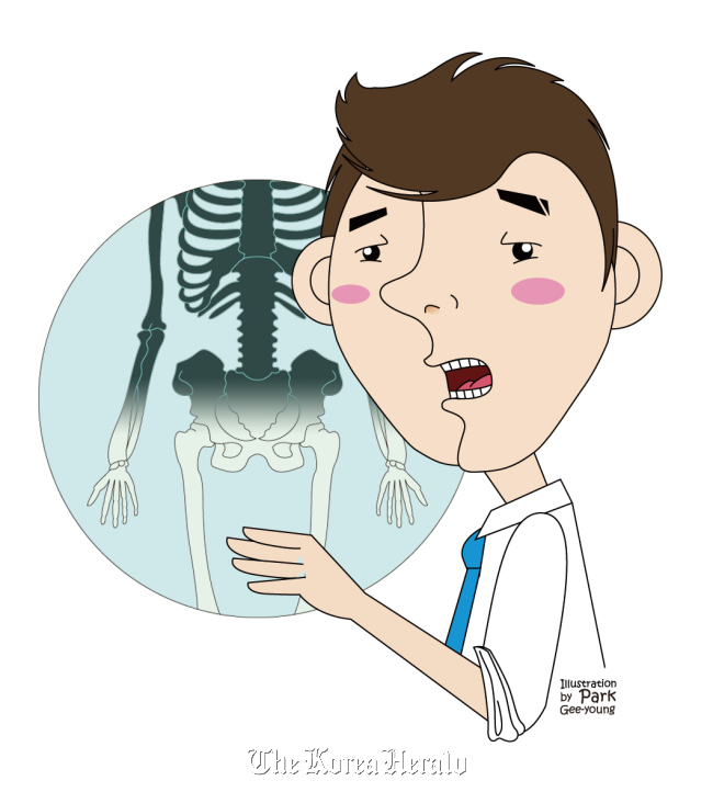
Our bones may seem sturdy and hard, but they are very active organs that undergo extensive synthesis and absorption. Bones are associated with growth, fracture repair and adjustment to exercise. We develop more bone in areas where they are needed more. For example, in environments with no gravity (as for astronauts in space), the bone becomes very weak and light. In athletes, the bones grow stronger because they are constantly under load.
Our bones are cylindrical in shape with an empty space in the middle. This allows the bones to resist forces. Bones consist of osteoblasts, which generate bones, and osteoclasts that break it down to calcium and other substances. The empty space in the middle of the bone is known as the medullary cavity and this is where stem cells are generated.
In osteopetrosis, the osteoblasts still have their normal function and continue to generate bone. However, the osteoclasts function abnormally and cannot break the bones down to calcium. This leads to the accumulation of excess bone made by the osteoblasts that cannot move out to other tissues, eventually causing the medullary cavities to fill with hard bone. Osteopetrosis is a rare condition.
Stem cells are made in the bone medullary cavities, which function as hematopoietic cells. Because the osteoclasts are dysfunctional, the bone medullary cavities fill with hard bone and the bones become more fragile. Such bones can fracture even with small forces (for example, from glass) and the decline in the number of platelets can eventually cause death.
Because there is defective bone resorption, the patients keep their bones from their childhood. The bones may look very dense and strong in X-rays, but they are often quite fragile and prone to fractures.
Congenital (childhood) Form: This form is inherited by an autosomal recessive form. Without treatment, the patients die within a few years. The disease manifests within the first 10 years of life.
Autosomal Dominant Form: Also known as marble bone disease, there is a systemic weakening and osteosclerosis of the bones. Recent studies consider this condition to be an autoimmune condition that involves a defect in the thymus, leading to the formation of abnormal osteoclasts.
Main symptoms
Congenital forms of this disease are characterized by thick, hard bone with poor modeling and narrow medullary cavities. The child often fails to thrive and shows variety of symptoms including myelophthisic anemia, thrombocytopenia, hepatosplenomegaly, lymphadenopathy, spontaneous contusion, abnormal bleeding and multiple fractures.
Abnormalities in the skull formation are also common. Due to abnormal bone formation, there is foraminal stenosis, which puts pressure on the cranial nerves. This can cause palsy of the optic nerve, occulomotor nerve and the facial nerve and can lead to blindness.
There are no reports of patients with congenital osteopetrosis who have survived more than 20 years without treatment. Patients have significantly defective immune systems and are prone to severe infections or bleeding to death.
Treatment
Fractures in these patients take a long time to heal. During the treatment of multiple fractures, there can be deformation of the coxa vara and the long bones, so corrective osteotomy is often needed. Dislocated pathological fractures should be treated using plate fixation as there is no bone cavity.
Osteomyelitis is common in this condition due to immune reaction following reduced blood supply. Therefore, it is important to monitor the patients closely after surgery.
Patients with the congenital form of this disease often die within the first two years of life. Those who develop the condition later in life survive to adolescence or adulthood. As there are frequent pathological fractures, patients should take care to avoid fractures. Children can also be treated with calcium or vitamin D.
 |
Park Youn-soo |
By Park Youn-soo
The author is a doctor at the Department of Orthopedic Surgery at Samsung Medical Center and a professor of Sungkyunkwan University school of Medicine. ― Ed.


